Science
AI-Enhanced Microscopy Offers Real-Time Views of Living Cells
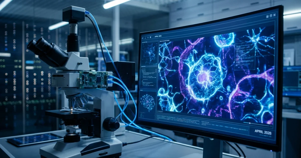
Artificial intelligence is rapidly transforming scientific imaging, as researchers now use AI-enhanced microscopy to capture crisp, real-time video inside living cells. According to Phys.org, these cutting-edge techniques are reshaping how scientists observe cellular processes, paving the way for advancements in disease research and treatment.
Sharper Images, Faster Results
Traditional microscopy methods have long struggled with limited resolution and slow imaging speeds, especially when observing dynamic processes within live cells. The integration of AI algorithms enables microscopes to achieve high-fidelity super-resolution imaging, producing clearer visuals at unprecedented speeds. This means researchers can now track cellular activity in real time, offering a window into processes previously too fast or too subtle to capture.
- AI-powered microscopy delivers crisp, detailed video of live cell interiors.
- Real-time imaging helps scientists monitor rapid cellular changes.
- Advanced algorithms reconstruct images from incomplete or noisy data, improving accuracy.
Transforming Disease Research
The ability to observe living cells as they interact and change offers new opportunities for understanding diseases at their root. By watching how cells respond to pathogens, drugs, or genetic mutations, researchers can identify early indicators of disease and test the effectiveness of potential treatments.
Phys.org notes that these breakthroughs could revolutionize fields such as cancer research, neurobiology, and infectious disease studies. Real-time imaging enables scientists to see how cellular processes unfold, rather than relying solely on snapshots at fixed points in time.
Key Benefits for Biomedical Science
- Earlier detection of abnormal cell behavior linked to disease.
- Improved screening of drug candidates by observing direct cellular responses.
- Enhanced understanding of disease mechanisms through dynamic visualization.
How AI Makes It Possible
AI-enhanced microscopy leverages deep learning models trained on vast datasets of cell images. These models can recognize patterns, reconstruct missing details, and distinguish subtle differences between healthy and diseased cells. For readers interested in the technical side, a comprehensive review of AI-powered live-cell imaging provides insights into the algorithms and comparative performance benchmarks.
By automating image interpretation, AI reduces the need for manual analysis and speeds up the discovery process. Researchers can now process large volumes of video data, extracting meaningful information far more efficiently than before.
Expanding Access and Collaboration
The impact of AI-enhanced microscopy extends beyond research labs. With open databases like the BioImage Archive, scientists worldwide can access and share high-quality microscopy datasets—both raw and AI-processed—accelerating collaborative projects and reproducibility.
Major funding agencies, including the NIH, are supporting development and deployment of advanced imaging tools, recognizing their potential to drive innovation in medical diagnostics and therapeutics.
Looking Ahead
AI-powered microscopy is still evolving, with ongoing research focused on refining algorithms, expanding imaging capabilities, and integrating with other technologies. As these methods mature, scientists anticipate even greater clarity and speed, unlocking deeper understanding of cellular life and disease.
Curious readers can explore the fundamentals in this advanced imaging primer to learn more about the science behind these innovations.
While challenges remain—including ensuring model accuracy and scaling technologies for widespread use—the promise of AI-enhanced microscopy is clear: real-time, high-resolution views inside living cells could transform how we diagnose, treat, and understand human disease.