Science
Johns Hopkins study reveals how the eye forms sharp central vision
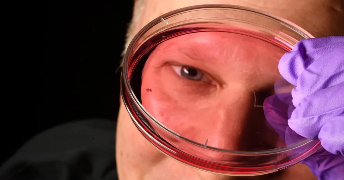
CYP26A1 and DIO2 are highly and continuously expressed in the central retina, where a Johns Hopkins team mapped a molecular switch that helps the human eye build its sharpest central vision before birth. The study challenges the long-held view that blue cone cells simply move away from the center of the retina and instead shows that cone fate can change under the right chemical signals.
The study, published Feb. 13 in the Proceedings of the National Academy of Sciences, focused on the central retina where two regulators, CYP26A1 and DIO2, are highly and continuously expressed. CYP26A1 breaks down retinoic acid, while DIO2 promotes thyroid hormone signaling, creating the conditions that steer cone-cell development in the foveolar center.
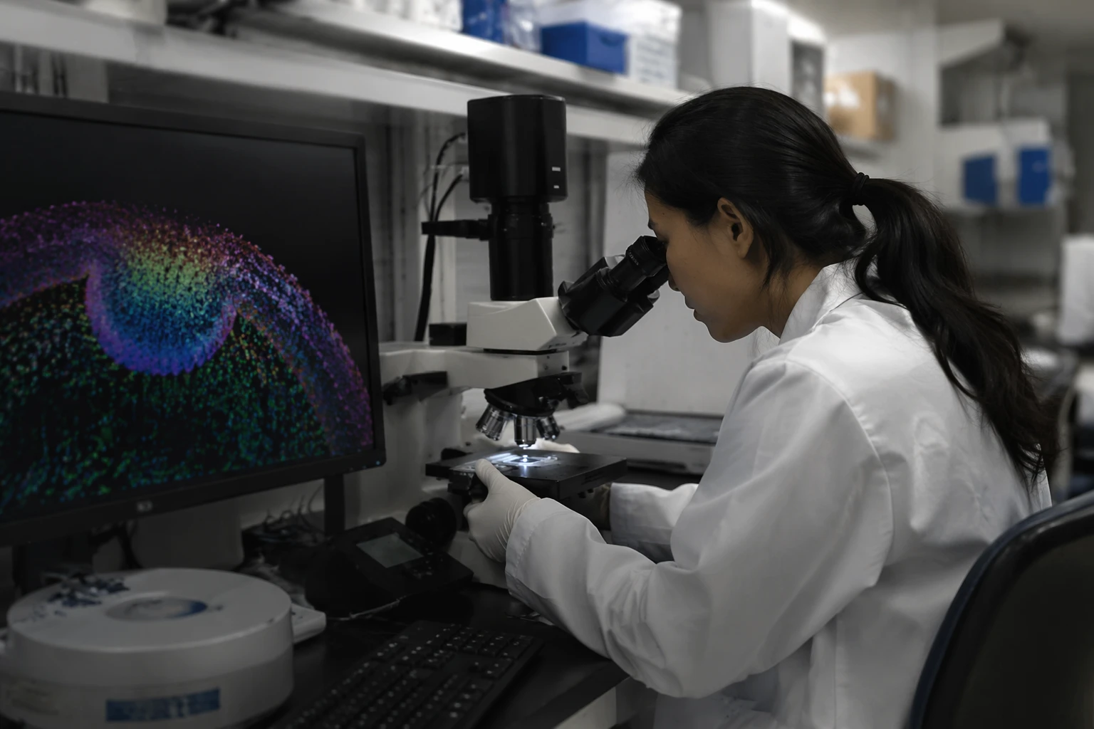
Researchers led by Robert J. Johnston Jr., an associate professor at Johns Hopkins, tracked development in human fetal retinas and retinal organoids over several months. In the organoid experiments, high retinoic-acid conditions increased S cones and reduced M/L cones. Sustained thyroid hormone signaling pushed cells toward M/L-opsin expression and even induced M/L opsin in cones that had already expressed S opsin, showing that cone identity is more flexible than once thought.
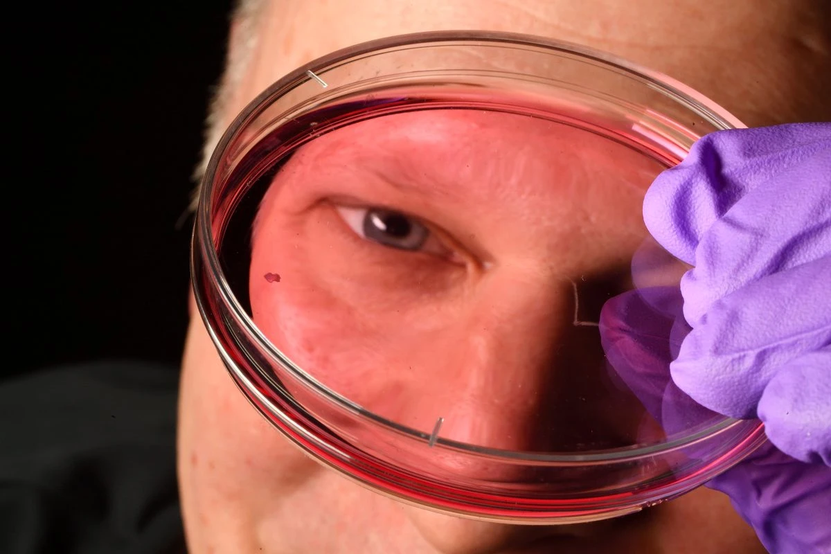
Inner retinal layers are displaced away from the center to reduce light scattering and make room for the dense cone packing that gives the foveola its high-acuity power. The biology is hard to study in animals because mice and fish do not share the same foveolar cone patterning. That has made human organoids, small tissue clusters grown from fetal cells, a valuable stand-in for the real tissue.