Technology
Simple microscopes get a billionfold boost, pinpoint amino acids
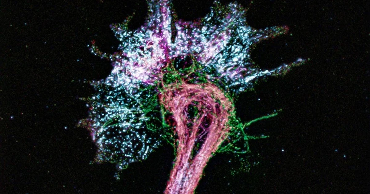
A Nature microscopy feature described a workaround that could move some of biology’s sharpest imaging out of elite facilities and into ordinary labs: stretch preserved protein samples in all directions until simple light microscopes can pinpoint amino acids. The journal framed the method as making samples one billion times bigger, a vivid shorthand for expansion microscopy, which physically enlarges tissue so structures that were once too small to see become visible with conventional optics.
Expansion microscopy began in Edward Boyden’s Massachusetts Institute of Technology lab in Cambridge in 2015, and the field has grown into a platform for super-resolution work on standard microscopes. The basic method embeds preserved specimens in a water-absorbing hydrogel, then swells the gel so biomolecules are pushed farther apart while their relative positions stay intact. That makes the biology appear larger without changing the microscope itself, which is why Nature described the technique in 2018 as a way to democratize super-resolution imaging.
The approach is no longer a niche proof of concept. The expansion microscopy community says more than 700 papers and preprints have now used it, and MIT has said the method has been applied to kidney disease, fruit fly brains, plant seeds, the microbiome, Alzheimer’s disease and viruses. In 2024, a Nature Methods paper reported single-shot 20-fold expansion microscopy, or 20ExM, which achieved isotropic roughly 20-times expansion of cells and tissues and under-20-nanometer resolution on a conventional microscope.
MIT News said in 2025 that new staining methods and image-processing tools let researchers see vivid outlines of cell shapes and pinpoint many different proteins inside a single tissue sample. The new Nature feature pushes that logic farther, toward amino acids themselves, with protein samples stretched enough that ordinary light microscopy can resolve details that once demanded much more expensive imaging systems. That matters not only for cell biology and structural biology, but also for diagnostics and research settings where access to top-tier equipment is limited.

The tradeoff is clear. Expansion microscopy is powerful for fixed samples, not live-cell imaging, so it cannot replace every method used to watch biology in motion. Even so, the technology is steadily lowering the cost of nanoscale observation, speeding up work, and widening access for labs that have long been locked out of the most advanced microscopes.